Published: October 2025 | Last updated: May 2026 | Editorial review and quality control: Martina N.
Finding a pale spot on the skin tends to start the same loop of questions: how long has that been there, did I miss something, is it contagious. Most of the time the answer is reassuring. White patches usually come from common causes like yeast that overgrows on warm skin, pigment cells that slow down after irritation heals, or a stable condition such as vitiligo, rather than from a sexually transmitted infection. This guide walks through what white patches typically mean, the cases that genuinely warrant a clinic visit or a home test, and how clinicians sort through the possibilities when someone walks in worried about a pale area they noticed in the mirror.
Key Takeaways
- Most white skin patches come from fungal overgrowth, vitiligo, healed irritation, or lichen sclerosus, not from a sexually transmitted infection.
- A patch becomes worth testing when it appears alongside sores, unusual discharge, pain, or follows a new sexual exposure.
What is actually happening when skin turns pale
Skin and mucous membranes only have so many ways to change color. When an area looks lighter than the tissue around it, one of three things is usually going on. The pigment-producing cells in that patch have slowed down or been destroyed, the surface of the skin has thickened so that it scatters light differently, or a layer of something else (most often yeast or a dermatophyte fungus, the category that causes ringworm and athlete's foot) is sitting on top of the skin and disrupting normal pigmentation.
That short list explains why a fungal infection and a pigment disorder can look almost identical from across the bathroom. Both produce pale areas. The differences show up in texture, in how the borders behave, in whether the patch itches or scales, and in whether it spreads or sits still for years. None of those clues is conclusive on its own, but together they shape what a clinician will check for first.
The other reason context matters: skin tone changes how patches present. On medium and darker complexions, even mild yeast or fungal involvement can leave noticeably pale spots because the affected area cannot pigment normally in response to sun. On lighter complexions, the same infection often reads as pink or tan rather than white.
| Mechanism | What is happening in the skin | Typical examples |
|---|---|---|
| Pigment cells slow down or die off | Melanocytes stop producing melanin in the affected area | Vitiligo, post-inflammatory hypopigmentation |
| Skin surface thickens | The outer layer becomes denser and scatters light differently | Leukoplakia, some scar tissue |
| Something is sitting on the skin | Yeast or fungus on the surface interferes with normal pigmentation | Tinea versicolor, cutaneous candidiasis, oral thrush |
When a white patch points to a fungal infection
Fungal overgrowth is one of the most common reasons skin develops pale or whitish areas, and it is treatable in most cases with antifungal creams, shampoos, or short courses of oral antifungal tablets (NHS, Ringworm). The fungi involved live on the outer layer of skin, feed on keratin, and thrive where it is warm and a little damp: the chest and back after summer workouts, the groin, under the breasts, and inside the mouth.
The classic presentation is tinea versicolor. The yeast that causes it (a Malassezia species) is part of normal skin flora and is not passed person-to-person through ordinary contact. It can overgrow during hot months or in people who sweat heavily, which is when the visible patches appear. As the yeast grows, it produces compounds that interfere with melanocyte function, leaving lighter patches that often become more obvious after sun exposure tans the surrounding skin (MedlinePlus, Tinea versicolor).
Cutaneous candidiasis behaves differently. The yeast prefers moist skin folds and tends to produce reddish patches with white satellite lesions at the edges, plus itching or burning. In the mouth, the same family of yeast causes oral thrush: creamy, cottage-cheese-style patches on the tongue or inner cheeks that can be gently scraped off to reveal raw, reddened tissue underneath (NHS, Oral thrush in adults).
| Condition | Where it appears | Typical signs |
|---|---|---|
| Tinea versicolor | Chest, back, shoulders, neck | Pale, pink, or tan patches with fine powdery scaling |
| Cutaneous candidiasis | Skin folds, groin, under breasts | Moist red patches with white satellite spots and itching |
| Oral thrush | Tongue, inner cheeks, palate | Creamy white patches that wipe off, leaving sore red tissue |
When the cause isn't fungus
One of the most common errors dermatologists see is months of antifungal cream applied to a patch that was never fungal in the first place. Several conditions look fungal at first glance and respond to nothing in the antifungal aisle.
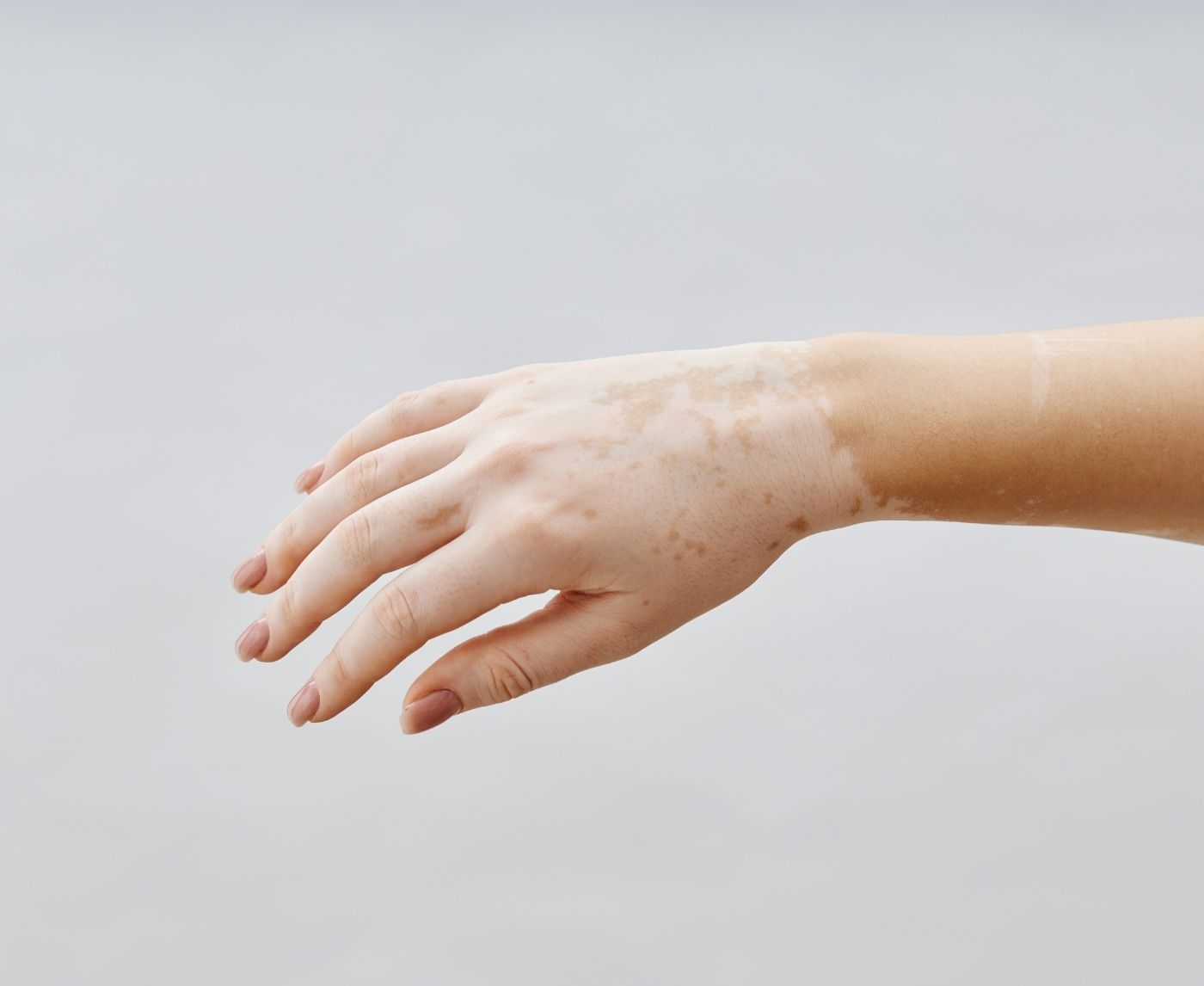
Vitiligo is the textbook example. It happens when the immune system targets and destroys melanocytes, the cells that give skin its color. The patches are usually sharply defined, completely white rather than slightly pale, and stable in shape for long periods. Vitiligo is not contagious, is not caused by hygiene, and tends to affect predictable areas like hands, around the mouth, and over joints (NHS, Vitiligo).
Lichen sclerosus is another non-fungal cause that shows up in clinics regularly, especially on genital and perianal skin. The patches are thin, shiny, slightly wrinkled, and can crack or itch. It is an inflammatory condition rather than an infection, and it usually responds to prescription steroid creams (NHS, Lichen sclerosus). Because it can look superficially like a yeast infection or like a mild trauma scar, people often delay getting it diagnosed.
Two more possibilities round out the differential. Oral leukoplakia produces thickened white patches inside the mouth that cannot be wiped away (unlike thrush). And post-inflammatory hypopigmentation is the lingering pale area left behind after eczema, acne, friction, or a healed rash. It is not infection at all, just pigment cells that have temporarily slowed down while the skin barrier recovers.
| Condition | What causes it | Clues it isn't fungus |
|---|---|---|
| Vitiligo | Autoimmune destruction of melanocytes | Sharply defined, completely white patches |
| Lichen sclerosus | Chronic inflammatory skin disorder | Thin, shiny skin, often in the genital area |
| Oral leukoplakia | Thickened oral tissue, often linked to irritation | White mouth patches that will not wipe off |
| Post-inflammatory hypopigmentation | Skin healing after rash or irritation | Lighter area appears after a known inflammation episode |
Where STIs fit into the picture
This is the question many readers came here to ask: can a white patch be a sign of a sexually transmitted infection. Yes, in specific situations a white patch can signal a sexually transmitted infection, but it is far less common than the first page of search results suggests. The article you are reading is published by stdrapidtestkits.com, which sells at-home STI testing kits; the relevant scenarios for testing are described below, and the rest of this section is about when a white patch genuinely warrants concern.
Syphilis is the clearest example. During the secondary stage of the disease, some people develop mucous patches: smooth, moist, gray-white lesions inside the mouth or on the genitals. These rarely appear in isolation. They typically come with a body rash (often on the palms and soles), swollen lymph nodes, and a recent history of unprotected exposure (CDC, About Syphilis).
HPV can produce small, light-colored or skin-toned bumps and plaques on genital tissue that some people mistake for fungal patches. These usually feel raised rather than flat and tend to cluster (WHO, STI fact sheet).
Persistent oral thrush in an adult who is otherwise healthy is occasionally the first sign of untreated HIV, because the immune system normally keeps Candida in check. Without other red flags, a single bout of thrush after antibiotics or steroid inhalers is far more likely to be a routine yeast event than an immune problem.
The takeaway: white patches in isolation, without sores, discharge, lymph node swelling, or a recent new exposure, are almost never an STI.
More than 1 million curable sexually transmitted infections (STIs) are acquired every day worldwide in people 15-49 years old, the majority of which are asymptomatic.
How clinicians actually tell the difference
Diagnosis rarely depends on appearance alone. White patches can look deceptively similar in the early days, especially before scaling or evolution sets in. A short sequence of low-effort tests usually narrows things down quickly, often inside one visit.
The first step is almost always a careful look at the patch under good light, checking for scaling, border character, distribution pattern, and whether the area changes when the surrounding skin is stretched. The next move depends on what the clinician sees. If fungal involvement is plausible, a gentle scrape of the patch onto a glass slide goes under the microscope, where Malassezia or Candida show up as recognizable patterns of hyphae (the thread-like strands that make up fungal cells) and budding yeast cells. A Wood's lamp examination (ultraviolet light) can highlight pigment loss and certain fungal organisms that fluoresce. When an STI is on the differential because of accompanying symptoms or exposure history, a swab or blood test gets added.
Most diagnoses are resolved by these steps. The cases that take longer are the ones where pigment loss looks like the early stage of something else, or where two conditions coexist (a yeast infection on top of long-standing post-inflammatory pigment change, for example). In those situations, dermatology follow-up is reasonable rather than starting empirical treatment.
| Step | Purpose |
|---|---|
| Skin examination under good light | Assess scaling, borders, distribution, and color patterns |
| Skin scraping for microscopy | Confirm or rule out fungal organisms within minutes |
| Wood's lamp examination | Highlight pigment changes and certain fungal species |
| Swab or blood test | Added when an infection such as an STI is on the differential |
What white patches usually look like in person
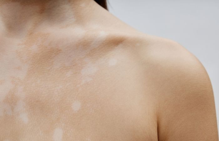
Most clinical-atlas photos online show advanced cases, which is part of why mild patches feel so alarming. In real life, the surface change is usually subtle: a slightly chalky area that you only notice once the surrounding skin has tanned, or a smoother section that looks marginally different from a few inches away.
When testing genuinely makes sense
Not every white patch needs a workup. Most can be watched for a couple of weeks, treated with an over-the-counter antifungal if the pattern fits, and reassessed if nothing changes. A few situations move things into the test-now category.
The clearest triggers are accompanying symptoms. A pale patch that comes with sores, painful urination, unusual discharge, bleeding, or swollen lymph nodes is no longer a stand-alone skin question. The same applies if the patch appeared shortly after sex with a new partner, especially without a barrier method, or if the affected area is on genital tissue and looks thin, shiny, or fragile (which can suggest lichen sclerosus rather than infection but still warrants evaluation).
Speed of change matters too. A patch that doubles in size within days, spreads to new areas of the body, or develops sores deserves a look. Patches that stay stable for weeks are usually less urgent. Persistent oral white patches that cannot be wiped off should also be checked, particularly in adults who smoke or drink heavily, because the differential there includes leukoplakia and not just thrush. Most leukoplakia patches are benign, but a small proportion can progress toward oral cancer over time, especially in heavy smokers and drinkers. That is why clinician evaluation rather than self-treatment is the appropriate response for persistent, non-wipeable white patches in those groups.
If the trigger is a recent sexual exposure rather than the patch itself, at-home rapid testing is reasonable while you wait for any other symptoms to clarify. Window periods matter (HIV antibody tests, for example, are most reliable from around 12 weeks after exposure per the CDC's HIV testing guidance), and many people find it less stressful to test once at the appropriate window than to refresh worst-case scenarios in their head daily.
A white patch moves out of the watch-and-wait category when any of these apply:
- Open sores, bleeding, or fragile-looking skin around the patch
- Unusual discharge or painful urination
- Swollen lymph nodes near the patch
- An oral white patch that will not wipe off, particularly in adults who smoke or drink heavily
- Appearance shortly after sex with a new partner without a barrier method
Why search results keep defaulting to fungus
Search engines reward the easy answer. Type "white patches on skin" and the first results almost universally jump to fungus, because fungal infections are the highest-volume cause and the easiest to explain. Volume and editorial simplicity get those articles ranked, even when the actual differential is broader.
There is a second issue with online imagery. Photographs that end up on medical sites tend to come from clinics, which means they document the moments when conditions had already progressed enough that someone sought care. Mild post-inflammatory hypopigmentation, faint early tinea versicolor, and the smooth pale patches of resolving eczema rarely get photographed. The visual library is skewed toward severity.
The result is predictable: someone scrolls for an hour, sees worst-case examples of each condition, and walks into the clinic convinced their faint patch is one of them. Clinicians commonly see patients who have either over-treated with antifungals for months or have catastrophized about cancer or HIV based on patches that turn out to be normal pigment variation.
The first failure mode is months of antifungal cream applied to a patch that was never fungal. The second is catastrophizing about cancer or HIV based on worst-case atlas photos when the real cause is mild pigment variation. Both delay accurate diagnosis and add anxiety that the patch itself rarely warrants.
Subtle clues your skin gives you
Pattern recognition is most of the work. Clinicians focus less on the color itself and more on how the patch behaves: whether it scales, whether the borders look sharp or fuzzy, whether it spreads outward from a center point, and whether the location fits a known cause.
Fungal patches tend to evolve. The center may become lighter or flaky while the borders expand outward in a roughly circular pattern. Vitiligo behaves differently. Its patches are smooth, sharply defined, and tend to stay stable for long periods, sometimes years, before any new spots appear. Lichen sclerosus often confines itself to genital and perianal skin and produces a thin, shiny surface that is uncomfortable rather than itchy in a fungal way.
Location adds the next layer of information. A patch on the chest or back after a sweaty summer points toward tinea versicolor far more often than anything else. A white patch on the tongue or inner cheek points toward thrush or leukoplakia. A pale plaque on genital skin opens up a broader list that includes lichen sclerosus, healed irritation, and (occasionally) syphilis or HPV.
| What you see | More likely explanation |
|---|---|
| Fine powdery scaling on chest or back | Tinea versicolor |
| Smooth, sharply defined completely white patch | Vitiligo |
| Thin, shiny, slightly wrinkled patch on genital skin | Lichen sclerosus |
| Creamy white patches in mouth that wipe off easily | Oral thrush |
| White patch alongside sores, discharge, or genital pain | Possible infection that warrants testing |
What happens after a diagnosis
If the patch turns out to be fungal, treatment is usually straightforward. Tinea versicolor responds well to topical antifungal creams or medicated shampoos used as body washes for one to two weeks. More widespread cases sometimes get a short course of oral antifungal medication. Cutaneous candidiasis clears with topical antifungals plus keeping the area dry. Oral thrush usually responds to antifungal lozenges or rinses (NHS, Oral thrush in adults).
A point that catches people out: pigment recovery takes longer than the infection. The fungus may be gone within days of finishing treatment, but the lighter spot can stay visible for weeks or even months while melanocytes ramp pigment production back up. That delayed recovery often gets misread as failed treatment.
For non-fungal causes, treatment depends on the underlying condition. Vitiligo management focuses on protecting affected skin from sun damage and (in some cases) topical immunomodulators to encourage repigmentation. Lichen sclerosus usually requires a prescription steroid cream, and untreated cases can lead to scarring, so it is worth not ignoring. Post-inflammatory hypopigmentation typically does not need treatment at all; gentle skin care, sun protection, and patience usually do the work.
The most important step is identifying the cause accurately. Antifungal creams on a pigment disorder do nothing useful, and steroid creams on an active fungal infection can make it worse.
What actually matters going forward
White patches almost always have an ordinary explanation. The combinations that genuinely warrant prompt evaluation are narrow, and a watch-and-wait approach handles the rest. If none of the red flags apply, a reasonable plan is to watch the area for a couple of weeks, try an over-the-counter antifungal if the pattern fits tinea versicolor or a simple yeast issue, and see what changes. If the patch is stable, painless, and not spreading, it is far more likely to be pigment-related than infectious. If it does evolve, a primary care visit or a dermatology referral usually clarifies things within one appointment.
If recent sexual exposure is part of the picture, at-home testing at the appropriate window period gives a clear result without a clinic appointment. For most people reading this, that result will be reassuring rather than alarming, and reassurance you have tested for is more durable than reassurance you have guessed for.
Triggers that move a patch from watch-and-wait to evaluation
- Rapid spreading or a patch that doubles in size within days
- Pain, open sores, bleeding, or fragile-looking skin around the patch
- Unusual discharge, painful urination, or swollen lymph nodes alongside the patch
- An oral white patch that will not wipe off, especially in adults who smoke or drink heavily
- Appearance shortly after sex with a new partner, particularly without a barrier method
FAQs
- Can a fungal infection actually make skin look white?
- Yes. Tinea versicolor is the classic example. The yeast that causes it produces compounds that suppress melanocyte function in the affected patch, so the area cannot pigment normally and reads as lighter than the surrounding skin. The effect becomes more visible after sun exposure tans the untouched areas.
- I noticed a pale patch after sex. Does that mean it is an STI?
- Usually not. Friction, sweat, lubricant residue, shaving, and condom material can all leave temporary skin reactions or pigment changes that look like a pale patch a day or two later. The combination that points toward an STI is the patch plus sores, unusual discharge, painful urination, or swollen lymph nodes, especially after exposure with a new partner.
- Why do fungal patches sometimes look lighter rather than red?
- Different fungi produce different reactions. Tinea versicolor disrupts pigment production rather than causing inflammation, which is why it tends to show up as pale or off-color rather than red. On darker skin tones the pale effect is more noticeable; on lighter tones the same infection may read as pink or tan.
- What is the easiest way to tell if a white patch is fungal?
- Clinicians look for fine powdery scaling and a slight stretch test. Gently scraping the surface of a tinea versicolor patch often produces a chalk-dust scale that vitiligo and post-inflammatory hypopigmentation do not. A microscope scraping or Wood's lamp examination confirms it within minutes if there is any doubt.
- Why do white patches sometimes appear after a rash heals?
- When skin recovers from inflammation (eczema, acne, friction, a bug bite, or even a healed scratch), the melanocytes in that area can temporarily slow down. The result is a lighter spot called post-inflammatory hypopigmentation. It can stay visible for several weeks to a few months before the normal color returns.
- What if the patch is in my mouth?
- White patches inside the mouth most often turn out to be oral thrush, especially after antibiotics, inhaled steroids, or illness. Thrush patches wipe off and leave a red surface. White patches that cannot be wiped off may be leukoplakia or an irritation-related thickening, and those should be evaluated by a clinician or dentist rather than treated with antifungals at home.
- How long should I wait before getting a patch checked?
- A practical rule: if a patch sticks around longer than three to four weeks, spreads to new areas, develops sores, or starts itching or hurting, get eyes on it. Minor skin changes usually fade within that timeframe. Persistent or evolving patches deserve a closer look, even if the most likely explanation is harmless.
- When does at-home STI testing make sense for someone worried about a white patch?
- Context matters more than the patch itself. If you have had a recent new sexual partner and the patch comes with other symptoms (sores, discharge, painful urination, swollen lymph nodes), testing is reasonable. Many people choose at-home rapid kits at the appropriate window period after exposure to replace daily worrying with a clear result, without a clinic appointment.
How we sourced this article: This guide is built from current public-health and dermatology guidance, including the CDC, the World Health Organization, the UK's National Health Service, and MedlinePlus. It does not replace a medical evaluation. For symptoms that concern you, see a licensed clinician.
- UK National Health Service. Ringworm overview, covering skin fungal infections including tinea and their treatment with antifungal creams and tablets.
- U.S. Centers for Disease Control and Prevention. About Syphilis page, describing the stages of disease including mucous patches in secondary syphilis.
- World Health Organization. Sexually Transmitted Infections fact sheet, with global incidence data on curable STIs and notes on asymptomatic infection.
- UK National Health Service. Vitiligo overview, including diagnostic approach and treatment options.
- UK National Health Service. Lichen sclerosus overview, covering presentation, diagnosis, and treatment with topical steroids.
- UK National Health Service. Oral thrush in adults, including the wipe-off test and risk factors such as antibiotics and inhaled steroids.
- MedlinePlus (U.S. National Library of Medicine). Tinea versicolor encyclopedia entry, including the role of Malassezia yeast in disrupting normal skin pigmentation.
- U.S. Centers for Disease Control and Prevention. HIV Testing guidance, including window-period information for the most common antibody tests.